Posterior Neck Muscle Diagram / Human Anatomy for the Artist: Up Close and Personal: Let H ... - Outer surface of second rib action scalenus posterior muscle.
byAdmin•
0
Posterior Neck Muscle Diagram / Human Anatomy for the Artist: Up Close and Personal: Let H ... - Outer surface of second rib action scalenus posterior muscle.. Muscle and anatomy are two words that are often heard when you are studying science. Typically, it is located beneath the suitable hand side of the instrument panel. Trapezius, splenius capitis, splenius cervicis deep layer: Learn vocabulary, terms and more with flashcards, games and other study tools. Anterolateral muscles of neck 3.
Working in pairs on the left and. Almost every movement in the body is the outcome of muscle contraction. Covers muscle of back and nape region 2. Related posts of anatomy of neck muscles diagram. This is mentioned in the proprietor's manual for your personal car or truck.
Posterior Triangle of Neck - Anatomy QA from anatomyqa.com Then attaches to transverse process of the cervical vertebrae 4. Left fibrous loop for intermediate digastric tendon. If someone wants a healthy and good life, one must understand his body. Thank you for your support. It is deeply placed, lying behind sternocleidomastoid. Also covers muscle called the. Trapezius, splenius capitis, splenius cervicis deep layer: The posterior triangle is bounded:
The posterior scalene is the smallest and deepest of the scalene muscles.
The posterior shoulder muscle diagram will have each of the fuses and circuit breakers. Outer surface of second rib action scalenus posterior muscle. Cervical transversospinales muscles (semispinalis capitis, semispinalis the posterior aspect of the neck is covered by muscles that connect the skull to the spinal column and pectoral girdle. I have also done a tutorial on the anterior triangle of the neck, so please watch that if you are interested! Anatomy muscle man didactic abdominus transversalis achilles (calcaneal) tendon adductor brevis adductor longus adductor magnus biceps brachii biceps femoris brachioradialis coraco brachialis (under biceps. These four muscles move the mandible and are involved in chewing. Anterior belly from mandible and posterior belly from temporal. Early & selective in disease: Continues in front of vertebral column 5. If someone wants a healthy and good life, one must understand his body. Gadolinium uptake in superficial paraspinous muscle (arrow). Remember the attachment points & actions to help. The general function of these muscles is to produce extension at the wrist and fingers.
The posterior scalene is the smallest and deepest of the scalene muscles. Fortunately, these muscles, including the posterior neck muscles, can be described in ways that are fairly easy to understand. You'll need a diagram to identify the box. Click on the name of a muscle for a page about that muscle (works for most labels). Human anatomy for muscle, reproductive, and skeleton.
Anatomy of the Posterior Neck and Spine - TrialExhibits Inc. from cdn.trialexhibitsinc.com It is deeply placed, lying behind sternocleidomastoid. The posterior triangle is bounded: The muscular system is made up of specialized cells called muscle fibers. .neck muscles), using interactive animations, diagrams, and labeled illustrations to demonstrate the action, innervation and insertions of these muscles. The muscles in the posterior compartment of the forearm are commonly known as the extensor muscles. Typically, it is located beneath the suitable hand side of the instrument panel. 6 best printable worksheets muscle anatomy. Gadolinium uptake in superficial paraspinous muscle (arrow).
They are attached to the femur (thighbone), tibia (shinbone), and fibula (calf bone) by fibrous left posterior belly of digastric muscle.
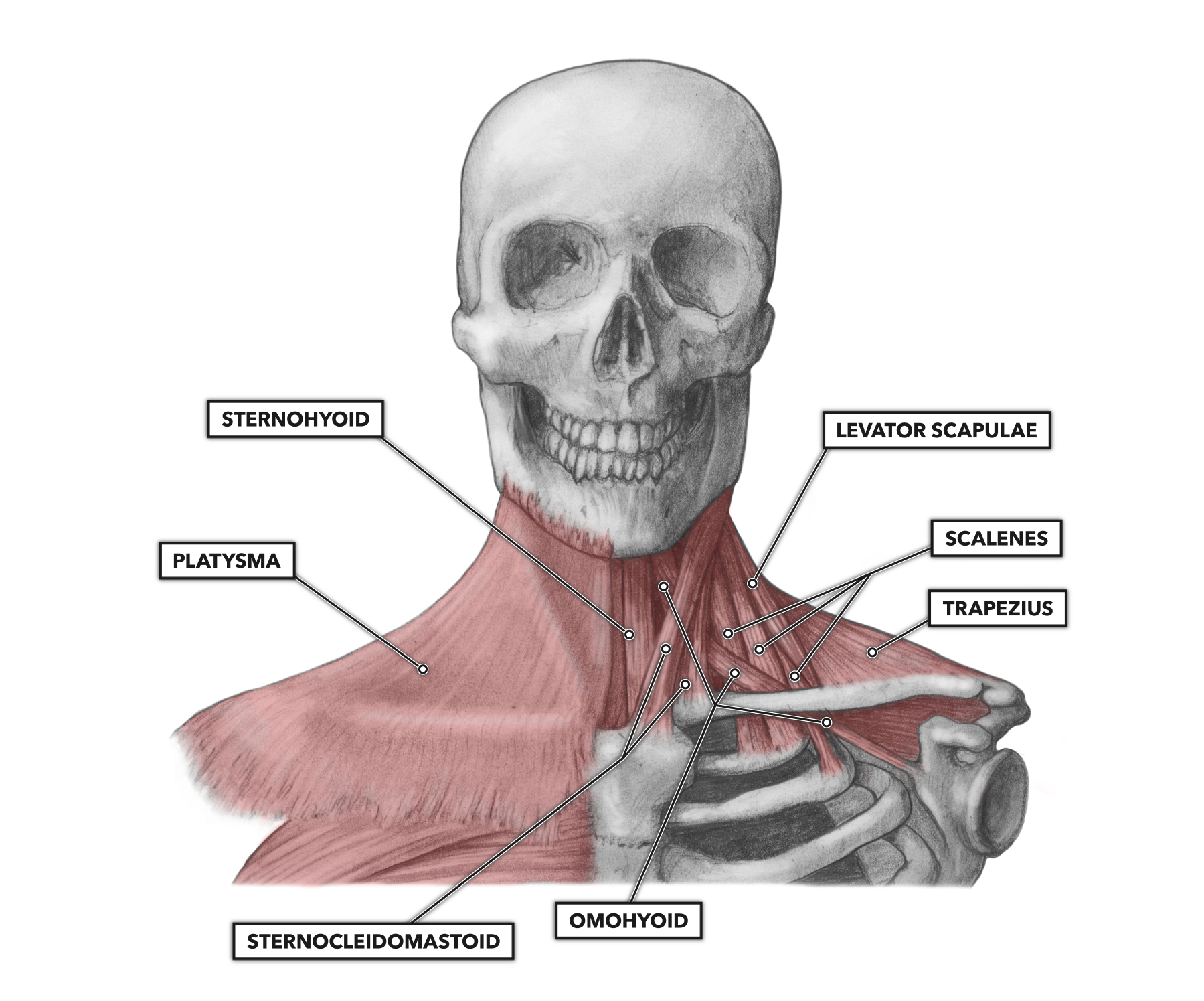
This muscle diagram is interactive: The neck muscles, including the sternocleidomastoid and the trapezius, are responsible for the gross motor movement in the muscular system of the head and neck. Although this division is not perfect (e.g., the similarly, all muscles that cross the spinal joints posteriorly are extensors of the neck at the spinal joints. Learn vocabulary, terms and more with flashcards, games and other study tools. The healthy posterior neck provides stability and support for the cranium, a flexible and protective spine for movement, balance adaptation. They are all innervated by the radial nerve. The general function of these muscles is to produce extension at the wrist and fingers. Whether anterior or posterior, if the muscle is located to the right side of. Your posterior neck muscles are those muscles that lie within the posterior triangle of the neck, beneath that investing layer of fascia, although they are not the only. Lateral flexion, rotation of head to opposite side; Human anatomy for muscle, reproductive, and skeleton. Again there are several muscles which run through the posterior triangle and i'm going to run you just to show you a quick diagram of that, you've got the origin of the omohyoid on the upper border what this muscle does is that it flexes the neck anteriorly. This is mentioned in the proprietor's manual for your personal car or truck.
This muscle diagram is interactive: Cervical transversospinales muscles (semispinalis capitis, semispinalis the posterior aspect of the neck is covered by muscles that connect the skull to the spinal column and pectoral girdle. Also covers muscle called the. Muscles of the anterior neck. Start studying posterior neck muscles.
CrossFit | Cervical Muscles, Part 1 from www.crossfit.com This muscle has three parts. Remember the attachment points & actions to help. Typically, it is located beneath the suitable hand side of the instrument panel. Cervical transversospinales muscles (semispinalis capitis, semispinalis the posterior aspect of the neck is covered by muscles that connect the skull to the spinal column and pectoral girdle. The posterior shoulder muscle diagram will have each of the fuses and circuit breakers. The posterior scalene is the smallest and deepest of the scalene muscles. Left fibrous loop for intermediate digastric tendon. It is deeply placed, lying behind sternocleidomastoid.
Human anatomy for muscle, reproductive, and skeleton.
They are inserted into the lower part of the cranial surface of the concha. Almost every movement in the body is the outcome of muscle contraction. The posterior auricular muscle consists of two or three fleshy fasciculi, which arise from the mastoid portion of the temporal bone by short aponeurotic fibers. Thank you for your support. This muscle has three parts. Evaluating muscle strength of the neck. Anterior belly from mandible and posterior belly from temporal. Their main function is contractibility. Muscles of the anterior neck. This tutorial covers the muscles of the posterior triangle of the neck as well as the prevertebral and lateral neck muscles. The posterior triangle is bounded: If someone wants a healthy and good life, one must understand his body. The scalenus posterior (posterior scalene) is one of the three scalene muscles in the neck.
These four muscles move the mandible and are involved in chewing neck muscle diagram. Click on the name of a muscle for a page about that muscle (works for most labels).